You are using an out of date browser. It may not display this or other websites correctly.
You should upgrade or use an alternative browser.
You should upgrade or use an alternative browser.
[تصاویر میکروسکوپی] بندپایان به روایت میکروسکوپ الکترونی
- شروع کننده موضوع Phyto
- تاریخ شروع


[FONT=century gothic, arial, helvetica][SIZE=-1]

The antennae, head and thorax of an aphid

[/SIZE][/FONT][SIZE=-1][FONT=century gothic, arial, helvetica]A view of the antenna and compound eye of an aphid[/FONT][/SIZE][FONT=century gothic, arial, helvetica][/FONT]
The antennae, head and thorax of an aphid
[/SIZE][/FONT][SIZE=-1][FONT=century gothic, arial, helvetica]A view of the antenna and compound eye of an aphid[/FONT][/SIZE][FONT=century gothic, arial, helvetica][/FONT]


شاخک سوسک Polposipus herculeanus
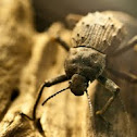

The antenna is covered in sensory hairs (blue) and cells (red), which are able to detect movement and smell. Beetles have two antennae positioned on their head between the eyes. The Fregate beetle is endemic to the Seychelles Island of Fregate, where it feeds on decaying trees. It has become critically endangered since the introduction of rats to the island
The antenna is covered in sensory hairs (blue) and cells (red), which are able to detect movement and smell. Beetles have two antennae positioned on their head between the eyes. The Fregate beetle is endemic to the Seychelles Island of Fregate, where it feeds on decaying trees. It has become critically endangered since the introduction of rats to the island

مگس تسه تسه
Coloured scanning electron micrograph (SEM) of a tsetse fly Glossina sp., a blood- sucking parasitic fly of tropical Africa. The fly pierces the skin of its host with the sharp probo- scis at lower right. The tsetse fly transmits Trypanosoma protozoa, of which T. gambiense & T. rhodesiense cause sleeping sickness in humans. Both male & female flies are bloodsuckers. Their habitat is varied, ranging from forest to river banks to savanna. Unlike most flies the tsetse gives birth to fully developed larvae, which immediately pupate. The female nurtures one larva at a time, with a total of 8-10 per lifetime. Magnification: x12.5 at 6x6cm size

Coloured scanning electron micrograph (SEM) of a tsetse fly Glossina sp., a blood- sucking parasitic fly of tropical Africa. The fly pierces the skin of its host with the sharp probo- scis at lower right. The tsetse fly transmits Trypanosoma protozoa, of which T. gambiense & T. rhodesiense cause sleeping sickness in humans. Both male & female flies are bloodsuckers. Their habitat is varied, ranging from forest to river banks to savanna. Unlike most flies the tsetse gives birth to fully developed larvae, which immediately pupate. The female nurtures one larva at a time, with a total of 8-10 per lifetime. Magnification: x12.5 at 6x6cm size
یک کنه که اینقدر خون خورده که داره میترکه!
The tick is swollen in size after feeding on the blood of its mammal host. The legs are protruding from its body either side of its mouthparts (centre) This is a common sheep tick (Ixodes ricinus), the principal vector of Lyme disease in Europe. It is common in the damp underbrush of European woods and attacks various domestic and wild animals, including dogs and humans. It carries the bacterium (Borrelia burgdorferi) that causes Lyme disease. Magnification: x18 when printed 10cm wide

The tick is swollen in size after feeding on the blood of its mammal host. The legs are protruding from its body either side of its mouthparts (centre) This is a common sheep tick (Ixodes ricinus), the principal vector of Lyme disease in Europe. It is common in the damp underbrush of European woods and attacks various domestic and wild animals, including dogs and humans. It carries the bacterium (Borrelia burgdorferi) that causes Lyme disease. Magnification: x18 when printed 10cm wide
لارو نوعی مگس از جنس Protophormia
The maggots of this fly are used medicinally to clean wounds. Its mouthparts are seen at centre right (grey). The maggots are sterilised and placed in the wound, where they feed on dead tissue and leave healthy tissue untouched. Their saliva contains anti- bacterial chemicals which maintain sterility in the area. Maggots are used on ulcers and deep wounds away from organs or body cavities, most often being used to treat diabetic ulcers on the feet. Magnification: x45 at 6x7cm size. x70 at 4x5"

The maggots of this fly are used medicinally to clean wounds. Its mouthparts are seen at centre right (grey). The maggots are sterilised and placed in the wound, where they feed on dead tissue and leave healthy tissue untouched. Their saliva contains anti- bacterial chemicals which maintain sterility in the area. Maggots are used on ulcers and deep wounds away from organs or body cavities, most often being used to treat diabetic ulcers on the feet. Magnification: x45 at 6x7cm size. x70 at 4x5"
کنه مخملی
Coloured scanning electron micrograph (SEM) of a velvet mite (Family Trombidiidae). Velvet mites are small predatory arachnids that live in the upper layers of soil. They are covered in fine hairs give them an appearance of velvet. Magnification: x40 when printed 10cm wide

Coloured scanning electron micrograph (SEM) of a velvet mite (Family Trombidiidae). Velvet mites are small predatory arachnids that live in the upper layers of soil. They are covered in fine hairs give them an appearance of velvet. Magnification: x40 when printed 10cm wide
نوعی مگس میوه
Its two compound eyes (red) are seen on either side of its head. At lower centre is its proboscis, used for sucking up food. This fly is widely used in genetic experiments as it breeds rapidly and has a small, well-understood genome (genetic code). Magnification: x90 at 6x7cm size. x140 at 4x5"

Its two compound eyes (red) are seen on either side of its head. At lower centre is its proboscis, used for sucking up food. This fly is widely used in genetic experiments as it breeds rapidly and has a small, well-understood genome (genetic code). Magnification: x90 at 6x7cm size. x140 at 4x5"
Similar threads
Similar threads
-

-
 [تصاویر میکروسکوپی]خیرهکنندهترین فراکتالهای طبیعی
- شروع شده توسط esmaeili60
- پاسخ ها: 14
-
 [تصاویر میکروسکوپی] تصاویری از دنیای گیاهان،جانواران و حشرات
- شروع شده توسط afsoon6282
- پاسخ ها: 77
-
 [تصاویر میکروسکوپی] مهاجمان ناپیدای زندگی ما
- شروع شده توسط maral22
- پاسخ ها: 20
-
{تصاویر طبیعت}حوض و آبگیری که به رنگهای مختلف در می آید
- شروع شده توسط afsoon6282
- پاسخ ها: 1